
مقال 13 دقيقة للقراءة
Is Endometriosis More Than One Disease?
New single-cell, spatial, and genetic research shows that lesions can have very different biology.
Endometriosis is not rare. It affects about one in ten women in their reproductive years. That is around 190 million women worldwide and 6.5 million in the U.S. alone. Yet in 2022, the NIH spent about $16 million on endometriosis research, or roughly $2 per patient. Compare that with Crohn’s disease, another chronic inflammatory condition that affects about a tenth as many Americans. It received $90 million in research funding that same year, or about $130 per patient.
You don’t need a calculator to see the problem. The math is not in endometriosis’s favor.
That funding gap is hard to ignore. It’s left clinicians without the tools they need, and women with endometriosis suffer. Pain gets dismissed as normal period cramps. A clear scan can end with “everything looks fine.” And there is still no reliable, noninvasive test that can definitively diagnose a condition affecting so many women
The last few years have started to change the story. Patient advocacy, grassroots momentum, and a broader push to close the gaps in women's-health research have helped move the field forward. We are now studying endometriosis lesions cell by cell and the results are challenging the idea that endometriosis is one uniform disease.
What “Tissue in the Wrong Place” Leaves Out
Endometriosis is often described as tissue similar to the uterine lining growing outside the uterus. That tells us where the disease is found, but much less about what a lesion is actually doing.
A lesion is made up of several kinds of cells. Some line glands. Others provide structure or help with the immune response. There are cells that form blood vessels and connective tissue, and some lesions even contain nerves. All of those cells respond to hormones, signals from one another, and the surrounding tissue.
Does where a lesion grows make a difference? An ovarian lesion is surrounded by different tissue than one on the pelvic lining or near the bowel. Its blood supply, immune activity, nearby nerves, and hormone signals can all be different. The question is whether those surroundings change the lesion itself. New research suggests they do.
Studying Endometriosis One Cell at a Time
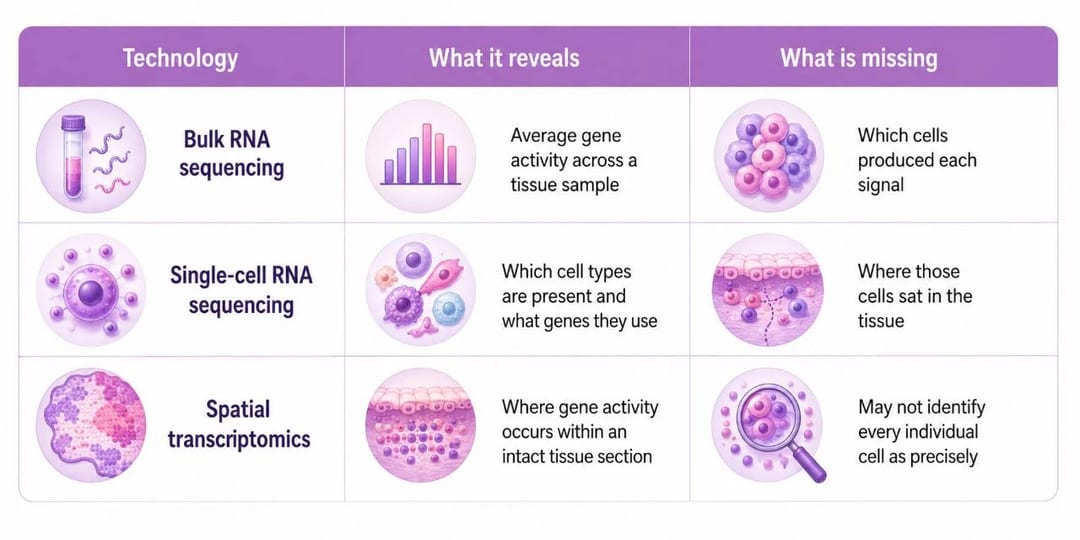
For years, most molecular-biology research relied on bulk RNA sequencing. In a typical experiment, researchers take a piece of tissue, extract all of its RNA, and measure which genes are active as an average across the whole sample. It is helpful, but it is a little like tossing a fruit salad in a blender and then trying to guess what fruit you started with.
If a lesion shows a strong inflammatory signal, bulk sequencing cannot tell us whether the sample contains more immune cells, whether those immune cells are unusually active, or whether another cell type is producing signals that attract them.
Single-cell RNA sequencing separates those signals by measuring gene activity in individual cells. It lets us identify the cell populations in a sample and see which genes are active in each one. The tradeoff is that, once the cells are pulled apart, we lose track of who was sitting next to whom in the tissue.
Single-cell RNA sequencing separates those signals by measuring gene activity in individual cells. It lets us identify the cell populations in a sample and see which genes are active in each one. The tradeoff is that, once the cells are pulled apart, we lose track of who was sitting next to whom in the tissue.
Spatial transcriptomics puts location back into the picture. Researchers can analyze a thin tissue slice and map gene activity across it. That lets us see where particular signals occur and which cells are close enough to influence one another. Think of a crowded dinner table. Bulk sequencing tells us how loud the room is. Single-cell sequencing tells us who is talking. Spatial transcriptomics shows us where everyone is sitting and which conversations are happening nearby.
If Figure 1 looks familiar, you are not imagining it. I covered these methods in my adenomyosis post, and the same tools are helping researchers ask similar questions in endometriosis.
Location Changes the Biology
Doctors already classify endometriosis by where it appears. Superficial peritoneal lesions are found on the thin lining of the pelvis and abdomen. Ovarian endometriomas are cyst-like growths on the ovary, often called “chocolate cysts” because old blood inside them turns dark. Deep endometriosis grows beneath the surface and can reach tissue near the bowel, bladder, or pelvic ligaments.
Those categories were based on clinical observations long before researchers could examine the underlying cell biology. A 2025 study published in Cell Genomics starts to fill in part of that picture. The researchers used both single-cell and spatial methods to examine 21 tissue samples, including ovarian endometriomas, deep endometriosis, and endometrial tissue from inside the uterus (eutopic) and outside it (ectopic).
Stromal cells are supportive cells in the uterine lining. Their gene activity normally shifts over the menstrual cycle. In the study, stromal cells from ectopic lesions retained part of that original cyclical identity while also showing changes associated with the environment in which the lesion was growing.
The ovarian endometriomas were especially interesting. Inside them, the researchers found distinct groups of stromal cells. Some had stronger inflammatory activity. Others showed features linked to fibrosis, the buildup of scar-like connective tissue. In some lesions, both patterns appeared in different areas. The ovarian tissue next to the lesion was changing too, with nearby cells showing disease-related changes of their own.
That does not mean every lesion follows the same pattern. It does suggest that a lesion and the tissue around it can influence one another. Ovarian endometriomas, superficial lesions, and deep lesions may share a diagnosis of endometriosis while existing in very different biological environments.
Hormone signaling may contribute to those differences. Endometriosis is often described as estrogen-dependent because estrogen helps lesion survive and grow. Progesterone ordinarily counterbalances some estrogen-driven effects, but cells in endometriosis can become less responsive to progesterone, a phenomenon often called progesterone resistance. Variation in local hormone signaling may help explain why lesions behave differently and why hormonal treatments work better for some women than others.
Why Lesion Severity and Pain May Not Match
The amount of visible disease does not always match the amount of pain. A small lesion in a highly inflamed area or near sensitive nerves can cause a lot of pain, while a larger patch somewhere else may cause very little. Lesions can trigger inflammation and repeated irritation. Fibrosis and adhesions can pull on nearby tissue, and nerves can grow around—or even inside—a lesion. A lesion near the bowel, bladder, or another nerve-rich area may cause very different symptoms from one in a less sensitive spot
When pain signals keep firing, they can also change how the nervous system handles pain. This is called central sensitization. Over time, it can make the nervous system more sensitive, which may help explain why pain can continue after surgery even when no visible disease remains. Lesion number and size matter, but they are not the whole story.
The Cells Inside a Lesion Are Talking to Each Other
Comparing lesions in different places is one way to study endometriosis. But if you look closely inside a single lesion, there is another kind of conversation happening between the cells.
A 2025 study in iScience used spatial transcriptomics to study superficial peritoneal lesions without breaking the tissue apart first. That meant the researchers could see which cells sat next to which. Burns and colleagues found evidence that epithelial cells lining the lesion may be signaling to nearby macrophages. Macrophages are immune cells involved in inflammation and tissue repair.
The next question was, who is the messenger? Complement component 3, or C3, stood out. C3 is part of the complement system, a group of proteins that helps defend against infection, clear debris, and regulate inflammation after tissue injury. Macrophages can respond to C3-related signals.
In normal wound healing, macrophages help clear debris and support repair before inflammatory activity settles down. But a lesion does not have an off switch, so inflammatory and repair signals may stay active longer than they would during normal wound healing. That kind of ongoing signaling can promote new blood vessel growth, encourage fibrosis, and make it easier for a lesion to persist.
The study didn’t directly demonstrate C3-mediated signaling between cells or prove that this pathway drives disease. Instead, the authors inferred potential communication from gene-expression patterns and cellular proximity. Even so, it shows the level of detail that spatial transcriptomics can reveal. Lesion epithelial cells and nearby immune cells may be participating in a local signaling network that helps sustain the lesion environment.
The Chemistry Inside a Lesion
RNA data reveal which genes are active, but they do not fully capture the biochemical activity inside a cell. Metabolism is the constant building, breaking down, and reshaping of molecules that keeps cells running. Metabolomics measures many of those small molecules, including lipids, amino acids, and sugars.
A 2025 study in Nature Communications combined single-cell sequencing, spatial transcriptomics, and spatial metabolomics to study ovarian endometriomas. Different areas within the same endometrioma showed different molecular features. The researchers also found links among inflammatory, fibrotic, and metabolic pathways in the local tissue environment.
These associations do not prove that one process directly causes the others. They do show that two parts of the same endometrioma can differ in cell composition, gene activity, and chemical state. RNA alone can’t fully measure how cells process nutrients, produce signaling molecules, or respond to metabolic stress. Those differences are not only transcriptional; they are biochemical as well.
The Changes May Extend Back to the Uterus
Most of what we know about endometriosis comes from studying lesions outside the uterus. A recent study in study in Nature Communications took a different approach, looking at the lining inside the uterus to see whether it changes in women with endometriosis.
The team analyzed endometrial biopsies from 35 women with endometriosis and 25 women without the condition, totaling more than 466,000 individual cells. Comparing the two groups revealed differences in gene activity across several cell types, including differences in cell-to-cell communication involving immune-related signals. The researchers also trained a machine-learning model on the single-cell data to distinguish endometriosis from control samples. The model had a median AUC of 0.83, which means it did a reasonably good job of separating the two groups in this dataset. This is encouraging, but it’s not the same as a clinically validated diagnostic accuracy measure, and the model still needs testing in larger, independent populations.
For now, the main point is that the endometrium inside the uterus can show disease-linked changes even when lesions are outside it.
Genetics Points to Some of the Same Biology
The studies above ask what is happening within lesions and the uterine lining. Genetics addresses a different part of the problem, the inherited susceptibility to developing endometriosis.
There is no single “endometriosis gene.” Risk comes from many genetic variants, each contributing a small amount. A 2026 Nature Genetics study examined nearly 1.4 million women, including about 106,000 with endometriosis. Koller and her team identified 80 regions of the genome linked to endometriosis risk, including 37 not previously reported. Five of those regions also overlapped with genetic risk for adenomyosis. Genetics cannot currently predict whether an individual woman will develop endometriosis, nor can it fully explain why one sister is affected and another is not. It can point scientists toward biological pathways worth investigating. When the researchers in this study matched inherited-risk signals with single-cell, spatial, and other molecular data, they highlighted the same biological processes involving immune regulation, hormone signaling, inflammation, tissue remodeling, and cell differentiation.
The methods differ in scale, from population-level DNA studies to molecular maps of individual lesions, but they are beginning to identify overlapping biology.
What’s Next
Single-cell and spatial studies are giving us a much more detailed view of endometriosis than we had only a few years ago. Most of this research has not reached the clinic yet, and none of it is a promise of a quick diagnostic test or a personalized treatment tomorrow.
The next step is to connect these molecular differences to what women actually experience. That means larger, more diverse studies that track lesion type, menstrual-cycle phase, medications, symptoms, treatment history, and outcomes. We also need to find out which differences are consistent enough to matter for individual women.
This is where readers, advocates, clinicians, researchers, and funders all have a role. Better studies need sustained investment, and better care will depend on earlier detection, and understanding why endometriosis can cause such different symptoms, lesion biology, and treatment responses from one woman to another. The emerging picture is not that endometriosis has suddenly become more complicated. It is that we can finally see more of the biology that was there all along, and begin asking which differences may matter for diagnosis, treatment, fertility, pain, and recurrence. #
References
- Liu S, et al. Single-cell and spatial transcriptomic profiling revealed niche interactions sustaining growth of endometriotic lesions. Cell Genomics. 2025;5(1):100737. doi
10.1016/j.xgen.2024.100737.
- Burns GW, Fu Z, Vegter EL, et al. Spatial transcriptomic analysis identifies epithelium-macrophage crosstalk in endometriotic lesions. iScience. 2025;28(2):111790. doi:10.1016/j.isci.2025.111790.
- Qi Y, Chen X, Zheng S, et al. Single-cell and spatially resolved omics reveal transcriptional and metabolic signatures of ovarian endometriomas. Nature Communications. 2025. doi:10.1038/s41467-025-66706-8.
- Dümpermann L, et al. Endometriosis-related alterations in the endometrium revealed by integrated single-cell and AI-powered approaches. Nature Communications. 2026. doi:10.1038/s41467-026-73020-4.